Overnight, Every Night!
What can time lapse microscopy do for your research?
- Generate compelling grant applications and supplemental data
- Get a new perspective on your biology
- Learn more faster and advance your research further

All Etaluma Microscopes can be used inside your incubator!

- Quality comparable to traditional, high-cost microscopes
- Compatible with microplates, dishes, chamber slides, flasks, microfluidic chips
- 3-Color fluorescence, phase contrast, brightfield
- Images, time-lapse & live video
- 1.25-100x objectives
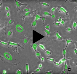
- LS720 pictured above with phase contrast: Auto XY stage & autofocus
"*" indicates required fields
LS850 Microscope
Blue, Green, & Red Fluorescence and Walk-Away Automation
Powerful, new LS850 Microscope is the latest generation of our fully automated three-channel flagship model and offers the latest advances in optics, cameras, throughput, and user flexibility. . The LS850 improves the image quality, motion speed, illumination, and software flexibility over the previous LS720 model. Exquisite XY motion control, motorized focus that allows autofocus and z-stacks, and easy-to-configure software combine to facilitate your automated microscopy experiments.
The LS850 in your incubator allows you to have a live cell imaging system that offers minimum photo toxicity and the most stable environment for long term imaging. Whether imaging multiple fields in your flasks or 1536 wells of cells with 3 fluorophores in a multi-day time-lapse, the LS850 offers a whole new world of walk-away automated microscopy!

LS820 Microscope
Blue, Green & Red Fluorescence with Manual Stage
The powerful, new LS820 Microscope is the latest generation of our fully automated three-channel motorized Z model and offers the latest advances in optics, camera, and user flexibility. The LS820 provides the image quality, Z-range, illumination, and software flexibility over the manual LS620 model. Motorized focus allows autofocus and z-stacks over 14 mm with 100 nm steps. Easy-to-configure software generating user readable and editable scripts, facilitate your automated microscopy experiments. The LS820 in your incubator allows you to have a live cell imaging system that offers minimum photo toxicity and the most stable environment for long term imaging. Whether imaging flasks, dishes or slides with 3 fluorophores in a multi-day time-lapse, the LS820 offers a compact solution for walk-away automated microscopy!

Features and Benefits
- Automated XY stage with autofocus in Z provides images (photos), time-lapse series, and videos recorded directly to your computer
- Fully functioning microscope empowers users to visualize cells from microplates, flasks, slides or custom labware
- Modern LED and advanced optical design provide near diffraction-limited (theoretical maximum) resolution
- Robust software allows set-up and control across many locations, including microplates and custom arrays
- Versatile and compact design enables use inside cell culture incubators and hoods
- Detects blue, green and red fluorophores, including BFP, DAPI, FITC, Fluo-4, GFP & mCherry
- Flip-up deck allows easy objective access
- Used manually, but also robot compatible (RS485, RS232, 5V digital interfaces)
- Objective compatibility with standard lenses permits use of your own objectives
Both utilize a Semrock Brightline® Pinkel filter set optimized for 405 nm, 488 nm, and 594 nm LED light sources. The 5-filter set is comprised of 3 exciting filters, 1 triple-band emitting filter, and 1 beam-splitting filter.
Excitation and Emission Profiles of LS850 and LS820 filters

| Comparison of Lumascope Models | ||
|---|---|---|
| Features | LS820 | LS850 |
| Optics | Blue, green & red fluorescence; bright field | Blue, green & red fluorescence; bright field |
| Phase Contrast | Phase contrast optional (Auraphase) | Phase contrast optional (Auraphase) |
| Objective Options | 1.25x,2.5x, 4x, 10x, 20x, 40x, 60x, and 100x(oil) magnification | 1.25x,2.5x, 4x, 10x, 20x, 40x, 60x, and 100x(oil) magnification |
| Objective Compatibilities | RMS-threaded, infinity corrected, 45 mm parfocal distance | RMS-threaded, infinity corrected, 45 mm parfocal distance |
| Fluorescence Filters | Blue: Ex 370-410, Em 429-462 nm; Green: Ex 473-491, Em 502-561 nm; Red: Ex 580-598, Em 612-680 nm | Blue: Ex 370-410, Em 429-462 nm; Green: Ex 473-491, Em 502-561 nm; Red: Ex 580-598, Em 612-680 nm |
| Camera | High Sensitivity Monochrome CMOS BSI Sensor; 5 MP 12-Bit | High Sensitivity Monochrome CMOS BSI Sensor; 5 MP 12-Bit |
| Image Formats | JPG, BMP, TIF, or PNG | JPG, BMP, TIF, or PNG |
| Image Size | Adjustable up to 2100 x 2100 pixels | Adjustable up to 2100 x 2100 pixels |
| Field of View | Up to 0.78 x 0.78 mm at 20x | Up to 0.78 x 0.78 mm at 20x |
| Video Rates | 25 FPS (exposure limited) | 25 FPS (exposure limited) |
| Stage | Manual XY | 120 mm x 80mm travel, 1-3 micron positional repeatability |
| Motorized Z | 14mm travel, 100nm step, image-based autofocus, Z-stacks | 14mm travel, 100nm step, image-based autofocus, Z-stacks |
| Well-to-well time (seconds) | NA | Image 96 wells: under 2 minutes with 1 channel, no auto-focus |
| Control Software | LumaviewPro, multi operating system application | LumaviewPro, multi operating system application |
| Computer Requirements | Windows 10, 11; Core i7, 512TB SSD, 8GB RAM MacOS with M1; 512TB SSD, 8GB RAM | Windows 10, 11; Core i7, 512TB SSD, 8GB RAM MacOS with M1; 512TB SSD, 8GB RAM |
| Integration | Python Source under the MIT Open Source License | Python Source under the MIT Open Source License |
| Power Requirements | USB3 for LS; 100-240 V, 50-60 Hz for autostage | USB3 for LS; 100-240 V, 50-60 Hz for autostage |
| Dimensions (WxDxH) | 24cm W x 22.6cm D x 27.8cm H (9.4in W x 8.9in D x 10.9in H) | 39.8cm W x 29.7cm D x 29.7cm H (15.6in W x 11.6in D x 11.6in H) |
| Weight | 5 kg (10 lb) | 9.5 kg (21 lb) |
| Operating Conditions | 0°C - 42°C, 5% - 95% RH non-condensing | 0°C - 42°C, 5% - 95% RH non-condensing |
Copyright © 2018 Etaluma, Inc. All rights reserved. Website design by Eskisse