Lumascopes
Applications
Featured Lumascope Applications
Lumascopes (fluorescence microscopes) can be used in practically any application where a standard inverted (and many upright) microscopes are used. Most of the applications below involve cells but Lumascopes can also image beads and other materials.
Time-lapse video of HT1080 cells expressing cell cycle proteins on an LS600; provided by Alex Zambon, Keck Graduate Institute (see Video Gallery for more info)
Overcoming the Physiological and Practical Constraints of Live Cell Imaging
A 2026 study from UC Santa Cruz, “A Modular In-Incubator Microscope for Longitudinal Live Cell Microscopy,” brings a critical conversation to the forefront of cell biology: the limitations of traditional imaging.
Learn how Etaluma’s Technology helped the team overcome these restraints
Widefield Fluorescent Imaging in Cell Morphology Studies
Discover how Lumascope’s high-contrast imaging supports detailed cell morphology analysis in fluorescence and brightfield modes. This article explores practical use cases in biological research and highlights the imaging power of widefield fluorescent microscopy.
Read the full case study on cell morphology imaging
Combining Phase Contrast and Fluorescence Microscopy
Learn how researchers enhance sample visibility by integrating phase contrast with fluorescence microscopy using Lumascope instruments. This approach enables non-invasive imaging of live cells while maintaining fluorescence signal clarity.
Explore the article on dual-modality microscopy
Microfluidic Microscopy
See how time-lapse microfluidic microscopy with the Etaluma LS720 captures 3D co-culture vessel and adipocyte formation over 7 days.
Read about our microfluidic capabilities here
Supporting Cancer Research with the Lumascope
Explore the advantages of Lumascope technology for enhanced imaging and analysis as was used for this exciting new cancer research paper.
Read the article and explore the advantages
Live Cell Bioluminescence Microscopy
Bioluminescence microscopy is gaining momentum as a low-light, live-cell imaging method. Etaluma’s new Lumi microscope is ready!
Live cell imaging
See Etaluma – Cardiac Myocytes Undergoing Division
Cell growth and confluence
See Time Lapse Video of MSC in 2D Cell Culture
Cell migration and wound healing
See Cell Migration & Wound Healing Application Note
See Migration of MSC in 2D Cell Culture
Cell cycle protein expression
See Human HT1080 Fibrosarcoma Cells with LS600
Use of micro-environmental systems
See Bioptechs products on Etaluma LS500
Calcium assays
GCAMP5 activity in a sensory neuron
Determining transfection efficiency
In Vitro Exercise Model
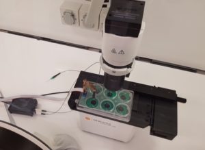 Cultured skeletal muscle myotubes are electrically stimulated under hypoxic conditions and with temperature manipulations. Cell signal transduction dynamics are measured using proteomic techniques to help understand how exercise stressors are translated into fitness-promoting adaptions such as increased mitochondria. Probe in photo measures PO2 in the cell medium rather than in the atmosphere. LS620 allows visualization of contracting cells and assessment of their health.
Thank you to Dr David Clarke and his lab, Laboratory for Quantitive Exercise Biology, Simon Fraser University, British Columbia, Canada
Behavior of stem cells
See Etaluma-Human Neural Stem Cells in Culture 1
See Etaluma-Human Neural Stem Cells in Culture 2
Also see reattachment of neuronal stem cells passaged with Accutase (scroll down to see video)
Cell death assays
Apoptosis induction
Spheroid development and behavior
See 3D Spheroid Formation of MSC
See Spheroid-Migration of MSC in a PEG-Fibrinogen Hydrogel
Cultivation of yeast
See Cultivation of S. cerevisiae in Core-Shell Microcapsules
Intravital studies
See Series: Neutrophil migration intravital mouse imaging
Study of lower eukaryotes
Copyright © Etaluma, Inc. All rights reserved.