Lumascopes
Applications: Microfluidic Microscopy and Co-Culture Complexity
“For the first time, experimental conditions capable of reproducibly forming diverse microvascular networks from telomerase immortalized endothelial and mesenchymal stem cells in both 2D and 3D hydrogel-embedded cultures are reported.”
Live Imaging of Vascular-Adipose Co-Culture in Microfluidic Systems
A recent study from the BMSE Lab at the University of Queensland, led by Mark Allenby, demonstrates a reproducible approach to forming stable microvascular networks alongside adipocyte differentiation within 3D hydrogel-based co-cultures. Utilizing telomerase-immortalized human endothelial and mesenchymal stem cells, the team has achieved concurrent vascularization and adipogenesis in a controlled microenvironment.
Central to the work is a microfluidic platform engineered to produce counter-current gradients of vasculogenic and adipogenic factors. This spatial arrangement supports the formation of physiologically relevant tissue structures and enables live-cell imaging throughout the culture period. The setup facilitates meaningful studies of vascular-adipose crosstalk by allowing precise spatiotemporal control over the culture conditions.
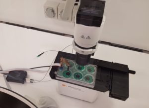
Time-lapse imaging was performed using the Etaluma LS720 microscope, capturing dynamic cellular behaviors over multiple days. (Note: the LS850 is Etaluma’s latest model.) At a 1:5 seeding ratio of endothelial to mesenchymal stem cells, networks remained adherent and viable through at least 7 days. Higher seeding ratios (2:1, 1:1) led to faster network formation but resulted in detachment, while lower ratios (1:2, 1:10) produced stable and branching structures.
The co-culture system also significantly enhanced adipogenic outcomes: lipid coverage reached 67% in co-culture, compared to under 2% in monoculture conditions. These results highlight the importance of engineered microenvironments in studying complex multicellular interactions.
Reference: Murphy, Franco, and Allenby, “Microfluidic Counter-Current Co-Culture to Model Adipose-Vascular Crosstalk,” Small (2025). https://onlinelibrary.wiley.com/doi/full/10.1002/smll.202501834
See Other Use Cases and Features of our Lumascopes
Live cell imaging
See Etaluma – Cardiac Myocytes Undergoing Division
Cell growth and confluence
See Time Lapse Video of MSC in 2D Cell Culture
Cell migration and wound healing
See Cell Migration & Wound Healing Application Note
See Migration of MSC in 2D Cell Culture
Cell cycle protein expression
See Human HT1080 Fibrosarcoma Cells with LS600
Use of micro-environmental systems
See Bioptechs products on Etaluma LS500
Calcium assays
GCAMP5 activity in a sensory neuron
Determining transfection efficiency
In Vitro Exercise Model
 Cultured skeletal muscle myotubes are electrically stimulated under hypoxic conditions and with temperature manipulations. Cell signal transduction dynamics are measured using proteomic techniques to help understand how exercise stressors are translated into fitness-promoting adaptions such as increased mitochondria. Probe in photo measures PO2 in the cell medium rather than in the atmosphere. LS620 allows visualization of contracting cells and assessment of their health.
Thank you to Dr David Clarke and his lab, Laboratory for Quantitive Exercise Biology, Simon Fraser University, British Columbia, Canada
Behavior of stem cells
See Etaluma-Human Neural Stem Cells in Culture 1
See Etaluma-Human Neural Stem Cells in Culture 2
Also see reattachment of neuronal stem cells passaged with Accutase (scroll down to see video)
Cell death assays
Apoptosis induction
Spheroid development and behavior
See 3D Spheroid Formation of MSC
See Spheroid-Migration of MSC in a PEG-Fibrinogen Hydrogel
Cultivation of yeast
See Cultivation of S. cerevisiae in Core-Shell Microcapsules
Intravital studies
See Series: Neutrophil migration intravital mouse imaging
Study of lower eukaryotes
Photomicroscopy in locations without AC power
Copyright © Etaluma, Inc. All rights reserved.