Lumascopes
Overcoming the Physiological and Practical Constraints of Live Cell Imaging
A significant 2026 study from UC Santa Cruz, “A Modular In-Incubator Microscope for Longitudinal Live Cell Microscopy,” brings a critical conversation to the forefront of cell biology: the limitations of traditional imaging.
The Hidden Cost of Environmental Disruption
The Ehrlich et al. paper identifies a “status quo” problem in many labs: the reliance on moving culture plates from an incubator to a standalone microscope. While this seems like a minor inconvenience, the biological impact is profound. Every trip out of the incubator subjects cells to:
- Thermal Shock: Even brief exposure to room temperature can alter metabolic rates.
- Gas Fluctuations: Changes in CO2 and O2 levels can trigger stress signaling pathways, particularly in sensitive hypoxia studies.
- Physical Agitation: Movement can disrupt delicate 3D structures, such as organoids or non-adherent cell clusters.
By utilizing an in-incubator system, researchers eliminate these variables, ensuring that the phenotypes observed are a result of the experiment, not the environment.
Engineering for Thermal Stability and Longitudinal Reliability
One of the most compelling aspects of the UCSC research is its focus on thermal stability. The researchers noted that heat-generating components (like high-power LEDs and electronics) must be managed carefully to prevent focus drift over multi-day experiments.
Etaluma Insight: This technical challenge is exactly why the Lumascope features a rugged, solid-state design. By minimizing moving parts and optimizing heat dissipation, our systems maintain focus throughout a 72-hour (or week-long) time-lapse. When your optics are designed to live at 37°C, you no longer have to worry about the “thermal expansion shimmy” that plagues traditional motorized microscopes.
Capturing the “Between-the-Frames” Biology
Traditional imaging is often limited by human schedules—checking cells at 9:00 AM and 5:00 PM. The UCSC paper demonstrates that longitudinal, automated imaging allows for the quantification of morphological remodeling in real-time. This is vital for:
- Transient Phenotypes: Identifying short-lived events, like a specific cell-to-cell interaction or a rapid apoptosis trigger.
- Kinetic Analysis: Moving beyond “start vs. end” data to see the velocity and trajectory of cell migration or wound healing.
- Modular Scalability: The ability to scan multiple plates or different experimental conditions simultaneously without manual intervention.
Future-Proofing for Microfluidics and Organ-on-a-Chip
Finally, the researchers emphasized modularity. As labs move toward complex models like Organ-on-a-Chip (OOC), the imaging system must accommodate fluidic tubing and perfusion pumps. An in-incubator microscope with a compact footprint allows researchers to build these complex “micro-environments” directly on the stage, turning a standard incubator into a sophisticated discovery platform.
Conclusion
The findings from UC Santa Cruz validate a clear trend: the future of cell biology is continuous, automated, and undisturbed. By integrating high-resolution optics directly into the physiological environment, we aren’t just taking better pictures; we are seeing more accurate science.
See Other Use Cases and Features of our Lumascopes
Live cell imaging
See Etaluma – Cardiac Myocytes Undergoing Division
Cell growth and confluence
See Time Lapse Video of MSC in 2D Cell Culture
Cell migration and wound healing
See Cell Migration & Wound Healing Application Note
See Migration of MSC in 2D Cell Culture
Cell cycle protein expression
See Human HT1080 Fibrosarcoma Cells with LS600
Use of micro-environmental systems
See Bioptechs products on Etaluma LS500
Calcium assays
GCAMP5 activity in a sensory neuron
Determining transfection efficiency
In Vitro Exercise Model
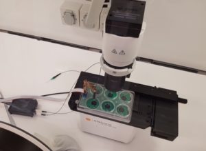 Cultured skeletal muscle myotubes are electrically stimulated under hypoxic conditions and with temperature manipulations. Cell signal transduction dynamics are measured using proteomic techniques to help understand how exercise stressors are translated into fitness-promoting adaptions such as increased mitochondria. Probe in photo measures PO2 in the cell medium rather than in the atmosphere. LS620 allows visualization of contracting cells and assessment of their health.
Thank you to Dr David Clarke and his lab, Laboratory for Quantitive Exercise Biology, Simon Fraser University, British Columbia, Canada
Behavior of stem cells
See Etaluma-Human Neural Stem Cells in Culture 1
See Etaluma-Human Neural Stem Cells in Culture 2
Also see reattachment of neuronal stem cells passaged with Accutase (scroll down to see video)
Cell death assays
Apoptosis induction
Spheroid development and behavior
See 3D Spheroid Formation of MSC
See Spheroid-Migration of MSC in a PEG-Fibrinogen Hydrogel
Cultivation of yeast
See Cultivation of S. cerevisiae in Core-Shell Microcapsules
Intravital studies
See Series: Neutrophil migration intravital mouse imaging
Study of lower eukaryotes
Photomicroscopy in locations without AC power
Copyright © 2024 Etaluma, Inc. All rights reserved.