Lumascopes
Applications: Phase Contrast and Fluorescence Microscopy
Analyzed to determine the percentage of EGFP-positive cells
Phase Contrast and Fluorescence in Uveal Melanoma Research
Uveal melanoma (UM) is the most common intraocular tumor in adults, yet researchers face a major challenge: the absence of commercially available normal choroidal melanocyte (NCM) cell lines for comparison. In a recent study titled “Co-isolation of human donor eye cells and development of oncogene-mutated melanocytes to study uveal melanoma,” researchers developed innovative methods to isolate and engineer donor eye cells to better understand how this cancer progresses.
To visualize these cells and their behavior in culture, the research team relied on phase contrast and fluorescence microscopy—two complementary imaging techniques that reveal structural and molecular details critical for cancer modeling.
Phase contrast microscopy allowed researchers to observe live, unstained cells in high detail, making it easier to track morphology and growth patterns over time. Meanwhile, fluorescence microscopy enabled the team to label and detect specific proteins and genetic markers related to oncogenic transformation, providing deeper insight into how healthy melanocytes become cancerous.
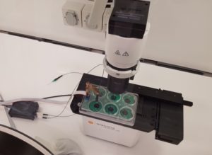
Etaluma’s compact and easy-to-use Lumascope microscopes were ideally suited for this research, enabling high-resolution imaging directly inside incubators or on the benchtop. The dual capabilities of phase contrast and fluorescence provided the flexibility needed to capture both structural and molecular changes in real time.
This combination of techniques supports more accurate disease modeling and may help pave the way for better screening, drug testing, and personalized therapies in the future.
Read the full research article here.
")
NCMs and RPE cells were successfully co-isolated and cultured. a An illustration of human eye anatomy and the location of RPE cells and NCMs. b Images of eye dissection and sequential RPE cell and NCM isolation. c Immunofluorescence analyses validated cell identity and purity. Scale bars = 30 μm
See Other Use Cases and Features of our Lumascopes
Live cell imaging
See Etaluma – Cardiac Myocytes Undergoing Division
Cell growth and confluence
See Time Lapse Video of MSC in 2D Cell Culture
Cell migration and wound healing
See Cell Migration & Wound Healing Application Note
See Migration of MSC in 2D Cell Culture
Cell cycle protein expression
See Human HT1080 Fibrosarcoma Cells with LS600
Use of micro-environmental systems
See Bioptechs products on Etaluma LS500
Calcium assays
GCAMP5 activity in a sensory neuron
Determining transfection efficiency
In Vitro Exercise Model
 Cultured skeletal muscle myotubes are electrically stimulated under hypoxic conditions and with temperature manipulations. Cell signal transduction dynamics are measured using proteomic techniques to help understand how exercise stressors are translated into fitness-promoting adaptions such as increased mitochondria. Probe in photo measures PO2 in the cell medium rather than in the atmosphere. LS620 allows visualization of contracting cells and assessment of their health.
Thank you to Dr David Clarke and his lab, Laboratory for Quantitive Exercise Biology, Simon Fraser University, British Columbia, Canada
Behavior of stem cells
See Etaluma-Human Neural Stem Cells in Culture 1
See Etaluma-Human Neural Stem Cells in Culture 2
Also see reattachment of neuronal stem cells passaged with Accutase (scroll down to see video)
Cell death assays
Apoptosis induction
Spheroid development and behavior
See 3D Spheroid Formation of MSC
See Spheroid-Migration of MSC in a PEG-Fibrinogen Hydrogel
Cultivation of yeast
See Cultivation of S. cerevisiae in Core-Shell Microcapsules
Intravital studies
See Series: Neutrophil migration intravital mouse imaging
Study of lower eukaryotes
Photomicroscopy in locations without AC power
Copyright © 2024 Etaluma, Inc. All rights reserved.