Lumascopes
Applications: Supporting Cancer Research with the Lumascope
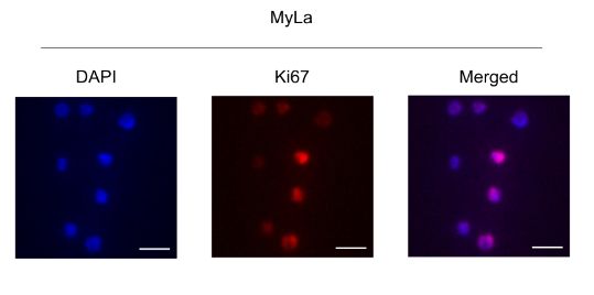
“To evaluate proliferation, we performed immunofluorescence staining for Ki67…Visualisation and photos were acquired with a Lumascope LS720 microscope”
Cutaneous T‐cell lymphoma (CTCL) Imaging with the Lumascope
 A recent peer-reviewed study led by researchers from McGill University and Harvard explored molecular factors involved in cutaneous T-cell lymphoma (CTCL), a rare form of skin cancer. Among the many tools used in the study, the Etaluma Lumascope played a key role in the researchers’ ability to visualize and quantify cellular activity through immunofluorescence imaging.
The team used the Lumascope to perform imaging of CTCL cell lines stained for Ki67, a marker commonly used to evaluate cell proliferation. By acquiring high-quality fluorescence images, the researchers were able to assess differences in proliferation between control and experimental groups where gene expression was altered. This helped them evaluate the potential impact of a gene called GTSF1 on cancer progression.
Though the study utilized the previous-generation Lumascope 720, today’s Lumascope 820 offers even greater imaging performance for live-cell and fixed-cell fluorescence applications.
Why Researchers Choose Lumascope
Etaluma’s Lumascope series continues to support academic and clinical researchers who need dependable, space-efficient imaging systems. In this CTCL study, the Lumascope allowed the team to:
- Capture detailed fluorescence images of cultured cells using minimal bench space
- Perform image-based quantification of proliferation using common nuclear markers
- Work within incubator conditions to maintain live or fixed samples
With intuitive software and no need for bulky components, the Lumascope line provides flexibility without compromising image quality. Its LED-based illumination and widefield optics make it a reliable option for users seeking consistent results across multi-day or high-throughput workflows.
The full article, including imaging data collected with the Lumascope, is available here.
See Other Use Cases and Features of our Lumascopes
Live cell imaging
See Etaluma – Cardiac Myocytes Undergoing Division
Cell growth and confluence
See Time Lapse Video of MSC in 2D Cell Culture
Cell migration and wound healing
See Cell Migration & Wound Healing Application Note
See Migration of MSC in 2D Cell Culture
Cell cycle protein expression
See Human HT1080 Fibrosarcoma Cells with LS600
Use of micro-environmental systems
See Bioptechs products on Etaluma LS500
Calcium assays
GCAMP5 activity in a sensory neuron
Determining transfection efficiency
In Vitro Exercise Model
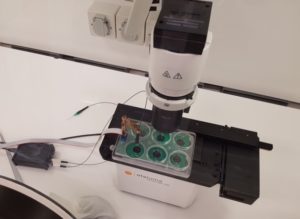 Cultured skeletal muscle myotubes are electrically stimulated under hypoxic conditions and with temperature manipulations. Cell signal transduction dynamics are measured using proteomic techniques to help understand how exercise stressors are translated into fitness-promoting adaptions such as increased mitochondria. Probe in photo measures PO2 in the cell medium rather than in the atmosphere. LS620 allows visualization of contracting cells and assessment of their health.
Thank you to Dr David Clarke and his lab, Laboratory for Quantitive Exercise Biology, Simon Fraser University, British Columbia, Canada
Behavior of stem cells
See Etaluma-Human Neural Stem Cells in Culture 1
See Etaluma-Human Neural Stem Cells in Culture 2
Also see reattachment of neuronal stem cells passaged with Accutase (scroll down to see video)
Cell death assays
Apoptosis induction
Spheroid development and behavior
See 3D Spheroid Formation of MSC
See Spheroid-Migration of MSC in a PEG-Fibrinogen Hydrogel
Cultivation of yeast
See Cultivation of S. cerevisiae in Core-Shell Microcapsules
Intravital studies
See Series: Neutrophil migration intravital mouse imaging
Study of lower eukaryotes
Photomicroscopy in locations without AC power
Copyright © Etaluma, Inc. All rights reserved.