Lumascopes
Applications: Watching Cancer Cells Squeeze Through Confined Spaces
More microfluidics, spheroids, and time-lapse — this time from researchers exploring how lung cancer cells migrate in tight environments.
Real Time Monitoring Made Possible Using Etaluma’s LS720 Microscope
In their recent paper Cancer Cells Traverse Faster in Confined Space by Modifying Vimentin Filaments With Nuclear Deformation and Promoting the Growth of Desired Tumor Spheroids (Alam et al., 2025), the team used a custom microfluidic platform to study how cells deform, reorganize, and adapt their cytoskeleton to move through narrow tracks.
What Researchers Found:
-
Cancer cells rely on softer, bleb-like protrusions when under spatial confinement, traveling faster and farther than in open channels.
-
These cells show nuclear deformation and shifts in vimentin filament organization — both key to squeezing through obstacles.
-
After migration, the same cells exhibited altered gene expression patterns, ultimately shaping how tumor spheroids form and grow.
Real-time monitoring of these protrusive behaviors was made possible using the Etaluma LS720 microscope, which allowed continuous imaging of migrating single cells in confined microchannels. (Note: the LS850 is Etaluma’s latest model.)The ability to track morphology and protrusion dynamics over hours of time-lapse was critical in linking cytoskeletal changes to spheroid development downstream.
Together, these results highlight the importance of microenvironmental stiffness and confinement in driving metastatic potential — and show how pairing microfluidic design with accessible live-cell imaging can uncover new dimensions of cancer biology.
Read the full research paper here.
See Other Use Cases and Features of our Lumascopes
Live cell imaging
See Etaluma – Cardiac Myocytes Undergoing Division
Cell growth and confluence
See Time Lapse Video of MSC in 2D Cell Culture
Cell migration and wound healing
See Cell Migration & Wound Healing Application Note
See Migration of MSC in 2D Cell Culture
Cell cycle protein expression
See Human HT1080 Fibrosarcoma Cells with LS600
Use of micro-environmental systems
See Bioptechs products on Etaluma LS500
Calcium assays
GCAMP5 activity in a sensory neuron
Determining transfection efficiency
In Vitro Exercise Model
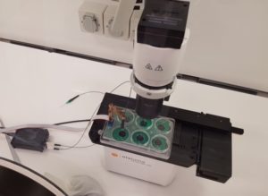 Cultured skeletal muscle myotubes are electrically stimulated under hypoxic conditions and with temperature manipulations. Cell signal transduction dynamics are measured using proteomic techniques to help understand how exercise stressors are translated into fitness-promoting adaptions such as increased mitochondria. Probe in photo measures PO2 in the cell medium rather than in the atmosphere. LS620 allows visualization of contracting cells and assessment of their health.
Thank you to Dr David Clarke and his lab, Laboratory for Quantitive Exercise Biology, Simon Fraser University, British Columbia, Canada
Behavior of stem cells
See Etaluma-Human Neural Stem Cells in Culture 1
See Etaluma-Human Neural Stem Cells in Culture 2
Also see reattachment of neuronal stem cells passaged with Accutase (scroll down to see video)
Cell death assays
Apoptosis induction
Spheroid development and behavior
See 3D Spheroid Formation of MSC
See Spheroid-Migration of MSC in a PEG-Fibrinogen Hydrogel
Cultivation of yeast
See Cultivation of S. cerevisiae in Core-Shell Microcapsules
Intravital studies
See Series: Neutrophil migration intravital mouse imaging
Study of lower eukaryotes
Photomicroscopy in locations without AC power
Copyright © Etaluma, Inc. All rights reserved.